Recently, the research team led by Associate Professor Tang Chuanxi from the Key Laboratory of Neurobiology of our school has made new progress in the research of lipid metabolism and the pathogenesis of Parkinson’s disease. Two research papers, namely “Enhanced Microglial Engulfment of Dopaminergic Synapses Induces Parkinson's Disease‐Related Executive Dysfunction in an Acute LPC Infusion Targeting the mPFC” and “Lysophosphatidylcholine Promoting α-Synuclein Aggregation in Parkinson’s Disease: Disrupting GCase Glycosylation and Lysosomal α-Synuclein Degradation”, have been published in Aging Cell (Top Journal in Q1 of Chinese Academy of Sciences Journal Partition) and npj Parkinson’s Disease (Top Journal in Q1 of Chinese Academy of Sciences Journal Partition) respectively. Xuzhou Medical University serves as both the first affiliation and corresponding institution for both studies.
Parkinson’s Disease (PD) is the second most prevalent neurodegenerative disorder after Alzheimer’s disease, mainly characterized by progressive loss of dopaminergic neurons in the substantia nigra pars compacta, which triggers motor symptoms such as tremor and myotonia. Nevertheless, non-motor symptoms including executive dysfunction (EDF) usually emerge prior to motor disorders, manifested as inattention, impaired decision-making and declined working memory, and effective intervention approaches have long been lacking. The prefrontal cortex (PFC) receives dopaminergic projections from the ventral tegmental area (VTA), and intact dopamine (DA) signaling in this region is critical for maintaining normal cognitive function. In addition, pathological aggregation of α-synuclein (α-syn) is one of the core events during PD progression. The role of abnormal lipid metabolism in PD has attracted growing research attention. Studies have shown that the level of lysophosphatidylcholine (LysoPC) is markedly elevated in serum and cerebrospinal fluid of PD patients and closely correlated with disease progression and severity, yet its impacts on the dopaminergic system and pathological proteins remain unclear. The team carried out systematic research focusing on the function of LysoPC in PD, revealing its pivotal regulatory mechanisms in neuroinflammation and disrupted pathological protein homeostasis, and offering novel insights into PD pathogenesis and potential intervention strategies.
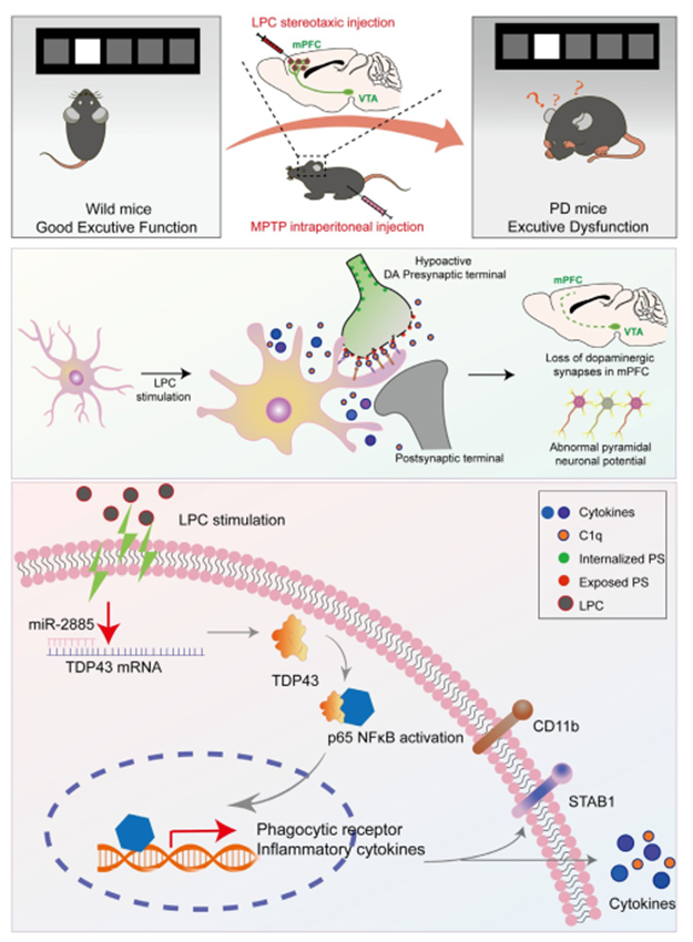
Finding 1 The study reveals that LysoPC triggers the miR-2885/TDP43/NF-κB signaling cascade to induce amoeboid activation of microglia. Activated microglia highly express Stabilin-1 and C3R, which recognize exposed "eat me" signals on dopaminergic terminals and lead to excessive engulfment of DA synapses. This pathological process reduces the excitability of glutamatergic neurons in the prefrontal cortex and ultimately results in executive dysfunction.
This research was jointly completed by Liu Yehao, postgraduate student majoring in neurobiology of our school, Chen Rui from Huai’an Affiliated Hospital of Xuzhou Medical University, and Mu Chunyan, doctoral candidate of neurobiology of our school as co-first authors; Associate Professor Tang Chuanxi of our school, Professor Zhang Zhi from Anhui Medical University and Director Qin Xiaoling from Xuhui Central Hospital, Fudan University as corresponding authors. The study was supported by the National Natural Science Foundation of China, Natural Science Foundation of Jiangsu Province, Talent Startup Fund of Xuzhou Medical University and Huai’an Natural Science and Technology Project.
Link to the original article: https://doi.org/10.1111/acel.70003
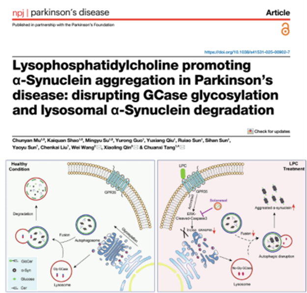
Finding 2 The study finds that within neurons, LysoPC activates the GPR35-ERK signaling pathway to induce Caspase-3 cleavage and suppress GRASP65 expression, thereby destroying the polarized structure of the Golgi apparatus, hindering glycosylation modification and lysosomal transport of GCase, and further causing accumulation of its substrate GlcCer, which acts as a scaffold for abnormal α-syn aggregation. Meanwhile, LysoPC exacerbates α-syn buildup by impairing autolysosomal function and lysosomal acidification, while solanesol can block the damaging effects mediated by ERK activation.
This research was jointly finished by Mu Chunyan (doctoral candidate in neurobiology), Shao Kaiquan (master’s student) and Su Mingyu (undergraduate of Class 2023, Major in Bioscience) of our school as co-first authors; Associate Professor Tang Chuanxi of our school, Dr. Wang Wei of Xuzhou Medical University and Director Qin Xiaoling from Xuhui Central Hospital, Fudan University as corresponding authors. The research received financial support from the National Natural Science Foundation of China, Jiangsu Youth Natural Science Foundation, Talent Research Fund of Xuzhou Medical University, Project of Hongkou District Health Commission of Shanghai, Jiangsu Graduate Student Innovation Fund and Student Science and Technology Innovation Project of Xuzhou Medical University.
Link to the original article: https://doi.org/10.1038/s41531-025-00902-7.
(First Reviewer: Dong Mengmeng; Second Reviewer: Gao Can; Third Reviewer: Li Chong)

